
|
Products
|


Tissue Simulation & Phantom Technology Model 711-HN
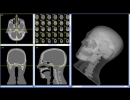
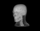
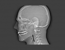
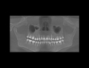
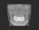
The CIRS ATOM Max Dental and Diagnostic Head Phantom is a standard of reference for diagnostic radiology of the head. The phantom is designed to assist technical and clinical staff in the selection, monitoring, training and verification of scanning parameters common to most radiological procedures requiring fine anatomical details.
The Model 711-HN provides a consistent tool for researchers, clinicians and technologists. It is ideal for determining optimum system settings, commissioning new equipment, monitoring system performance and training in dental X-ray, panoramic X-ray, CT and cone beam CT procedures.
The phantom includes an adjustable stand for positioning within a cone beam CT or panoramic X-ray system. The jaw of the phantom is slightly opened and front teeth are vertically aligned to replicate correct positioning with a bite guide. Please note that an actual bite guide cannot be positioned in this product.
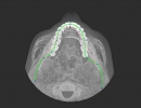
The phantom is constructed of proprietary tissue equivalent materials. ATOM®Max is made of tissue simulating resins that mimic the X-ray attenuation properties of human tissue for both CT and therapy energy ranges (50 keV-25 MeV). The Model 711-HN approximates the average male human head in both size and structure. The phantom includes detailed 3D anthropomorphic anatomy including brain, bone, larynx, trachea, sinus, nasal cavities and teeth. The bones contain both cortical and trabecular separation. The teeth include distinct dentine, enamel and root structure including the nerve. The sinus cavities are fully open.
Features:
- Includes detailed anatomical features
- Frankfurt plane identified to ensure proper alignment
- Tissue Equivalent from 50 keV to 25 MeV
- Positioning stand with six degrees-of-freedom
- Carrying case included
- 4 year warranty
Capabilities & Applications
- Commission X-ray, panoramic X-ray, CT and cone beam CT systems
- Learn how to properly position head for optimal images
- Test reconstruction techniques and algorithms for implant planning and maxillofacial reconstruction
- Train and evaluate personnel during implementation of new equipment and techniques
- Validate consistency of image quality
- Custom configurations are available upon request.
|
Call Us:
+65 69090758 Email:
|